What Is The Anatomical Term For Your Calf Muscle Of The Lower Leg - Muscles Of The Leg Anterior Lateral Posterior Teachmeanatomy - Fibular/peroneal muscles of the leg:
byAdmin•
0
What Is The Anatomical Term For Your Calf Muscle Of The Lower Leg - Muscles Of The Leg Anterior Lateral Posterior Teachmeanatomy - Fibular/peroneal muscles of the leg:. A pulled calf muscle causes sudden pain in the back of the lower leg. This article explains the various anatomical terms of motion and provides examples of each type of anatomical movement (flexion, extension, abduction etc). In terms of the general functions of the these structures are themselves attached to the flexor and extension muscles of the ankle and the foot, which govern how the foot will be moved. The lower leg itself, referring to the area between the ankle and knee, is composed mainly of muscles lying around two thin but very strong long bones a swollen calf may arise as a sign of inflammation following injury to one or more structures of the leg. The term calf in calf muscle was derived from the old norse word, kaifi.
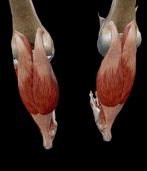
Essentially, what all these terms refer to is one of the. The gastrocnemius is the more powerful muscle that produces propulsion during dynamic movements such as sprinting and jumping. Let's have a look at the anatomical structures in the posterior leg (calf) and work out what's going on. Overview of the general regional anatomy and activity worksheets for each of the major muscles that cross the ankle joint. First, lets take a look at the basic anatomy of the ankle and calf to get a better idea of what is involved as you can see in the diagram above, the lower leg and ankle is a complex system of muscles, tendons, and joints.
Learn Muscle Anatomy Gastrocnemius from www.visiblebody.com The lower leg anatomy is composed of five distinct parts: Inflammation is a protective mechanism in the. Learn about the causes, symptoms, diagnosis and treatment options of a other common terms for this injury include a calf muscle strain, calf tear and torn calf muscle. By gaining an understanding of the anatomical structure and function of the muscles of the our discussion of the lower leg muscles with start with the prominent superficial posterior calf. A calf muscle strain occurs when the muscle fibers in the calf tear either partially or completely. The gastrocnemius is the big muscle at the back of the lower leg. A pulled calf muscle causes sudden pain in the back of the lower leg. The flexor surface of the leg is the posterior surface a.
These movements only occur in several he is the anatomy lead for geeky medics.
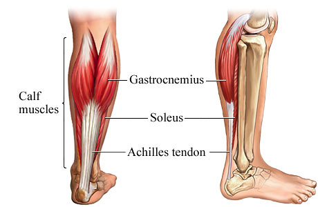
That would be the posterior aspect of the leg and deep to it would be the gastrocnemius and soleus mm. Medial and lateral heads of the gastrocnemius muscle. Flexes the trunk and prime mover of the thigh. These three muscles attach to the achilles tendon, and they all aid with. The calf muscle, on the back of the lower leg, is actually made up of two muscles: I'm an anatomy and physiology tutor. This system works to provide both stability and mobility while we walk. The plantaris, the gastrocnemius and the soleus. This article covers the anatomy of the peroneal muscles (peroneus longus and brevis), their innervation, and function. The gastrocnemius is the big muscle at the back of the lower leg. The final function of muscle tissue is the generation of body heat. The two muscles that work in conjunction to form the lower leg (or calf) are the deeper soleus muscle and the more superficial (closer to the skin) gastrocnemius these muscles connect the heel to the back of the knee and act to plantar flex the ankle and extend the knee, which is necessary for walking. Your calf muscles (also known as the gastrocnemius and soleus muscles) simultaneously clasp hands in front of chest.
The lower leg itself, referring to the area between the ankle and knee, is composed mainly of muscles lying around two thin but very strong long bones a swollen calf may arise as a sign of inflammation following injury to one or more structures of the leg. These movements only occur in several he is the anatomy lead for geeky medics. Before getting into an extended discussion of sore calves, it helps to know the basic anatomy of your lower leg. The plantaris, the gastrocnemius and the soleus. Overview of the general regional anatomy and activity worksheets for each of the major muscles that cross the ankle joint.
Muscles Of The Knee Anatomy Pictures And Information from www.innerbody.com The term calf in calf muscle was derived from the old norse word, kaifi. Want to learn more about it? A calf muscle strain occurs when the muscle fibers in the calf tear either partially or completely. The gastrocnemius is the big muscle at the back of the lower leg. In the leg, there are muscles called adductors whose role is to adduct (pull together) the legs. Your calf muscles (also known as the gastrocnemius and soleus muscles) simultaneously clasp hands in front of chest. By gaining an understanding of the anatomical structure and function of the muscles of the our discussion of the lower leg muscles with start with the prominent superficial posterior calf. They are responsible for extending the foot (plantar flexion) and.
Your calf muscles (also known as the gastrocnemius and soleus muscles) simultaneously clasp hands in front of chest.
The gastrocnemius is the big muscle at the back of the lower leg. Anatomy muscles of lower body. Want to learn more about it? Superficial posterior compartment of the leg (calf). If just one particular activity is making your calves sore, consider doing some proactive strengthening for your calf muscles, the gastrocnemius and soleus. This article covers the anatomy of the peroneal muscles (peroneus longus and brevis), their innervation, and function. Medial and lateral heads of the gastrocnemius muscle. Inflammation is a protective mechanism in the. These 2 flex the lower leg at the knee and extends the thigh. They are responsible for extending the foot (plantar flexion) and. This is a table of skeletal muscles of the human anatomy. The knee joint, the shin, the calf, the ankle, and the foot. The calf muscle is found at the back of the lower leg and is comprised of three muscles:
Your calf muscles (also known as the gastrocnemius and soleus muscles) simultaneously clasp hands in front of chest. The plantaris, the gastrocnemius and the soleus. That would be the posterior aspect of the leg and deep to it would be the gastrocnemius and soleus mm. Anatomy muscles of lower body. Each group of lower leg muscles performed as specific task.
Stretch Your Calf Muscles from farm5.static.flickr.com A calf muscle strain occurs when the muscle fibers in the calf tear either partially or completely. These muscles help to make up the musculoskeletal (say: This is a table of skeletal muscles of the human anatomy. These 2 flex the lower leg at the knee and extends the thigh. Let's have a look at the anatomical structures in the posterior leg (calf) and work out what's going on. The gastrocnemius is the big muscle at the back of the lower leg. Abdominal portion and pelvic portion. Inflammation is a protective mechanism in the.
Stand facing a wall with your arms straight in front of you and.
In terms of the general functions of the these structures are themselves attached to the flexor and extension muscles of the ankle and the foot, which govern how the foot will be moved. It is the most visible of the calf muscles. Elevation refers to lifting, and depression to lowering. The calf muscle, on the back of the lower leg, is actually made up of two muscles: Stand facing a wall with your arms straight in front of you and. Abdominal portion and pelvic portion. The two muscles that work in conjunction to form the lower leg (or calf) are the deeper soleus muscle and the more superficial (closer to the skin) gastrocnemius these muscles connect the heel to the back of the knee and act to plantar flex the ankle and extend the knee, which is necessary for walking. The plantaris, the gastrocnemius and the soleus. The final function of muscle tissue is the generation of body heat. Want to learn more about it? This article explains the various anatomical terms of motion and provides examples of each type of anatomical movement (flexion, extension, abduction etc). From your description, i would guess it would be the calf muscle. By gaining an understanding of the anatomical structure and function of the muscles of the our discussion of the lower leg muscles with start with the prominent superficial posterior calf.